Años 30

KERATOPLASTY: EXPERIMENTAL STUDY UPON THE EYES OF RABBITS WITH LEUKOMATOUS CORNEAS

Título: Keratoplasty: experimental study upon the eyes of rabbits with leukomatous corneas

Película en 35 mm, blanco y negro, sin audio, textos en inglés

Año de filmación: 1932

Duración: 11m 02s

Formato digital: WMV

Ubicación: Biblioteca del Instituto de Investigaciones Oftalmológicas Ramón Castroviejo

Signatura: IRC V-272

Esta película se proyectó en el XIV Concilium Ophthalmologicum (1933) y se complementa con la ponencia del Dr. Castroviejo "Queratoplastia: estudio experimental en conejos con córneas leucomatosas".

La comunicación sobre la película aparece recogida en el T. III del XIV Concilium Ophthalmologicum, publicado en Madrid en 1934.

Ver registro en Cisne

Descripción de textos incluidos:
0:53 The leucoma is produced by burning the eye with lime
1:22 Instruments required for the operation
1:36 A special keratome: 4 millimeters wide
1:45 Special scissors: the points are dull to avoid injury to iris and lens
1:55 Combination needle holder, scissors and forceps
2:04 Three months later, the inflammation disappeared, the rabbit is ready for the corneal transplant. The rabbit is again anaesthetized with Pernostan. The pupil must be fully dilated with atropine (3%), 15 minutes before the operation three instillations of epinephrine (1/1000), 5 minutes apart-are made to obtain an ischaemic conjunctiva.
2:28 The hair around the eye is clipperd as short as possible. Eyelids and adjoining areas are painted with iodine
2:52 A new drops of argyrols (20%) epinephrine (1/1000) and cocaine (4%9 are instilled into the eye
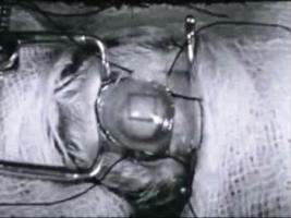
3:10 Speculum is inserted
3:16 Upper and lower conjunctival flaps are dissected and stretched to cover the central corneal area. Two sutures, abouth 5 millimeters apart, are placed reaching from the lower to the upper conjunctivl flap
4:00 Before proceeding with the operation one must be dure that the conjunctival flaps cover the center of the cornea, where the transplant is to be made.
4:16 Cross-incisions are made at right angles to each other with the keratoplastotome, the object being to outline a corneal flap 4 millimeters square. These incisions do not penetrate into the anterior chamber..
4:56 The incisions become sharply outlined after the instillation of fluoresceine"
5:05 While the eye is immobilized by taking hold of the episclera near the limbus-a puncture is madeat the upper incision with the keratome held at an angle of 45º"
5:19 The other three edges are cut with the special scissors held at an angle of 45º. Holding the keratome and scissors at an angle of abouth 45º results in uniformly bevelled edges of the flap
5:58 Similar technique is employed in obtaining the transparent flap from an enuceated eye
7:27 The transparent flap replaces the dissected leucoma
7:56 The sutures are tied and additional sutures are placed: usually one more on each side of the original two suffice to cover the transplant completely with the conjunctiva"
9:52 Metaphen ointment is applied, eyelids closed and dressing of cotton satured with collodion is placed, giving satisfactory protection
10:22 Six day later sutures are removed and a second dressing is applied, remaining for anther six days"
10:35 Transparent transplant more than one year old


KERATOPLASTY

Título: Keratoplasty

Película en 35 mm, blanco y negro, sin audio, textos en inglés

Año de filmación: 1937

Duración: 10m 33s

Formato digital: WMV

Ubicación: Biblioteca del Instituto de Investigaciones Oftalmológicas Ramón Castroviejo

Signatura: IRC V-273

Ver registro en Cisne

00: 29 - It is better top perform the operation under local, but this picture was taken of a nine year old child under general anaesthesia.Special instruments required for the operation.Keratoplastotome or double bladed knife
00:55 - Special keratome
01:02 - Special scissors with dull points to avoid injury to lens
01:13 - The pupil must be fully dilated with atropine 3%. A few drops of argyrol 20%, , epinephrine (1:1000), and pantocaine (2%) ae instilled into the eye. If local anaesthesia is employed, repeated instillations of pantocaine (1/2%) and adrenalin (1/2%) are used for about half an hour previous to the operation.
01:43 - A sub-conjunctival injection of cocaine (2%) and adrenalin (1:1000) produces a wider dilation of the pupil and reinforces the anaesthesia.
02:02 - Cross-incisions are made at right angles to each other with the keratoplastotome, the object being to outline a corneal flap. The incisions become sharply outlined after instilation of fluoresceine
02:44 - With fine corneal needles and nº 00000 silk a continuous suture is placed in the cornea alongside the incision
04:52 - A suture is placed in the center of the outlined area to hold the cornea away from the lens, while the leucoma is being dissected
5:11 - While the eye is immobilized by taking hold of the episclera near the limbus, a puncture is made at the upper incision with the keratome
05:35 - The other three edges are cut with the special scissors. Holding the keratome and the scissors at an angle of about 20 from the vertical, results in uniformly bevelled edges of the flap
06:31 - Similar technique is employed in obtaining the transparent flap from an eye enucleated shortly after delivery from a full-term still born. The transplant may also be obtained from eyes enucleated from adults
07:38 - The transparent flap replaces the dissected leucoma