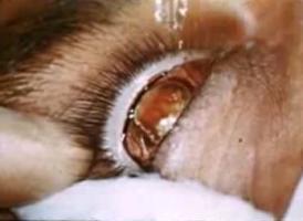
KERATOPLASTY (TREPHINE TECHNIQUE)
Ficha
Título
KERATOPLASTY (TREPHINE TECHNIQUE)
Descripción
Título: Keratoplasty (Trephine technique)
Película en 35 mm, color, sin audio, textos en inglés
Año de filmación: 1937
Duración: 10min 18s
Formato: WMV
Ubicación: Biblioteca del Instituto de Investigaciones Oftalmológicas Ramón Castroviejo
Signatura: IRC V-282
Ver película (Complumedia)
Descripción de textos incluidos:
00:30 The operation may be performed under local or general anaesthesia, although local is to be preferred. The pupil must be fully dilated with atropine 3%. A few drops of argyrol (20%), epinephrine (1:1000), and pantocaine (1%) are instilled into eye. If local anaesthesia is employed repeated instillations of pantocaine 1/2% and adrenalin 1:1000 are used for abouth half an hour previous to the operation
1:14 With the aid of a special trephine, 4 1/2 mm in diameter, an incision is made in the leucomatous cornea, the object being to outline a circular corneal flap. The incision becomes sharply outlined after instillation of fluoresceine.
1:46 With very fine atraumatic needles and Nº 00000000 (eight 0) black silk a continuous suture is placed in the cornea around the incision.
3:31 A suture is placed at the center of the outline area, wich will be used to hold the cornea away from the lens while the leucoma is being dissected.
3:52 The trephine is now used to complete the corneal incision, entering the anterior chamber, the central suture placed in the leucomatous outlined area having previously been passed within the hollow trephine.
5:00 If there is some part of theleucomatous disc still uncut, the dissection is finished with the aid of special curved scissors.
5:28 With the same trephine used in the eye of the host, a transparent flap is obtained from an eye enucleated shortly after delivery from a full term stillborn or from eye enucleated from adults on account of conditions which leave the conea unimpaired.
6:33 The transparent flap replaces the dissected leucoma
6:45 The corneal suture is tied
7:38 Metaphen ointment and a binocular dressing are applied.
7:52 Improvement of vision from hand motion to finger at abouth one foot to abouht 20/100 to 20/20
Catálogo Cisne
